Skull X-Ray
Skull X-Ray: Complete Guide to the Human Skull
What Is a Skull X-Ray?
A skull X-ray is a medical imaging examination that uses a small amount of X-ray radiation to create images of the bones of the skull. It can show the shape, position, and general condition of the skull and facial bones.
The skull protects the brain and supports important structures of the face. Because bone absorbs more X-rays than most surrounding soft tissues, the bones appear clearly on an X-ray image.
What Does a Skull X-Ray Show?
A skull X-ray can demonstrate many of the major bones and bony structures of the head.
Depending on the view taken, an image may show:
- Frontal bone
- Parietal bones
- Temporal bones
- Occipital bone
- Sphenoid bone
- Ethmoid bone
- Nasal bones
- Zygomatic bones
- Maxilla
- Mandible
- Eye sockets (orbits)
- Nasal cavity
- Paranasal sinus regions
- Sutures between skull bones
The exact structures visible depend on the X-ray projection and positioning.
Major Parts of the Skull
Frontal Bone
The frontal bone forms the forehead and contributes to the roof of the eye sockets. It is one of the major bones of the cranium.
Parietal Bones
There are two parietal bones, one on each side of the skull. They form a large portion of the upper sides and roof of the cranium.
Temporal Bones
The temporal bones are located on the sides of the skull near the ears. They contain important structures associated with hearing and balance.
Occipital Bone
The occipital bone forms much of the back and base of the skull. It contains the foramen magnum, a large opening through which the spinal cord connects with the brain.
Sphenoid Bone
The sphenoid is a complex bone located near the center of the skull. It contributes to the floor of the cranial cavity and the structure around the eye sockets.
Ethmoid Bone
The ethmoid bone is located between the eyes and contributes to the nasal cavity and the medial walls of the orbits.
Facial Bones
The skull also contains several facial bones, including the nasal bones, maxilla, zygomatic bones, and mandible.
How Does a Skull X-Ray Work?
X-ray imaging works by passing X-rays through the head toward a detector.
Different tissues absorb X-rays by different amounts. Dense bone absorbs more X-rays and therefore appears relatively bright on the resulting image, while areas that allow more X-rays to pass through appear darker.
The resulting image provides a two-dimensional representation of the structures captured by that particular projection.
Common Skull X-Ray Views
Different projections can be used to examine different parts of the skull.
Lateral View
A lateral projection produces a side view of the skull and can demonstrate the cranial bones, facial structures, and other anatomical landmarks.
AP or PA View
Anteroposterior (AP) and posteroanterior (PA) projections provide front-to-back views of the skull.
Towne View
The Towne projection can provide a view of the occipital region and other structures at the back of the skull.
The specific projections used depend on the clinical purpose of the examination.
Why Is a Skull X-Ray Performed?
Historically, skull X-rays have been used for evaluating certain bone abnormalities, fractures, foreign objects, and other conditions involving the skull.
Today, many head and brain-related situations are evaluated with other imaging technologies, such as CT, because CT can provide much more detailed cross-sectional information.
The appropriate imaging method depends on the clinical situation and the healthcare professional’s assessment.
Skull X-Ray vs CT Scan
A skull X-ray produces a relatively simple two-dimensional image.
A CT scan uses multiple X-ray measurements to create cross-sectional images and can provide considerably more detailed information about the skull and surrounding structures.
In situations where detailed assessment of a head injury or complex bone structure is required, CT may be preferred.
Interesting Facts About the Skull
- The adult human skull contains 22 bones when the auditory ossicles are excluded.
- The skull consists of the cranium and the facial skeleton.
- Skull bones are connected by immovable joints called sutures.
- The mandible is the largest and strongest bone of the facial skeleton.
- The skull protects the brain and provides attachment points for many muscles.
- The skull also contains openings called foramina, through which nerves and blood vessels pass.
What You Can See in Our X-Ray Image
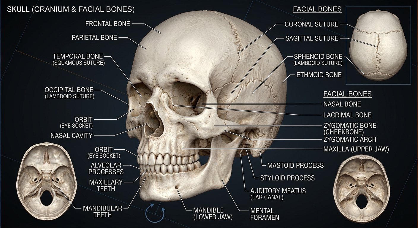
The TECH X-RAY image above illustrates important structures of the skull, including parts of the cranium and facial skeleton.
Use the labels in the image to locate structures such as:
Frontal Bone → Parietal Bone → Temporal Bone → Occipital Bone → Sphenoid Bone → Ethmoid Bone → Nasal Bone → Zygomatic Bone → Orbit
This makes the image useful as a visual anatomy reference.
| Feature | Details |
|---|---|
| Subject | Skull X-Ray |
| Category | Human Body |
| Imaging method | X-ray radiography |
| Main structures | Skull and facial bones |
| Image type | Two-dimensional projection |
| Related technology | CT scanning |
| Primary purpose | Visualization of bony structures |
Important Note
This article is provided for educational and informational purposes. An X-ray image should be interpreted by a qualified healthcare professional when used for medical diagnosis or treatment
Quick Information
Subject: Skull X-Ray
Category: Human Body
Published: August 27, 2026
Related X-Rays