Hand X-Ray
Hand X-Ray: What Can You See Inside the Human Hand?
The human hand is a remarkable structure made up of 27 bones, numerous joints, ligaments, tendons, muscles, nerves and blood vessels. A hand X-ray allows us to see the bones and their alignment from inside the body.
A normal hand X-ray can help students understand the arrangement of the bones, while healthcare professionals use X-rays as an important imaging method for evaluating injuries and abnormalities.
🩻 What Is a Hand X-Ray?
A hand X-ray is a medical imaging examination that uses a small amount of X-ray radiation to create images of the bones and joints of the hand.
Because bones absorb more X-rays than soft tissues, they appear relatively bright on an X-ray image. This makes the technique particularly useful for examining the skeletal structure.
🦴 How Many Bones Are in the Hand?
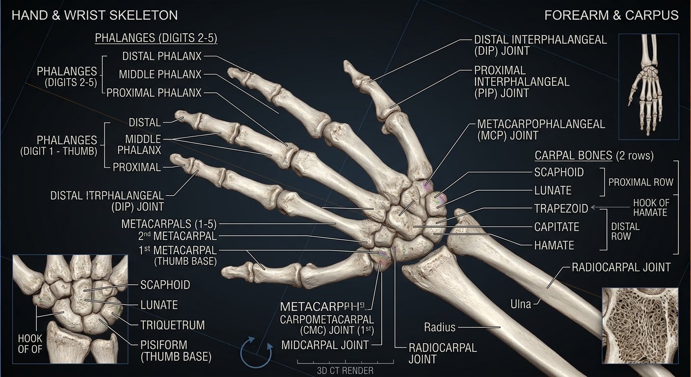
A human hand contains 27 bones:
- 8 Carpal bones — wrist
- 5 Metacarpal bones — palm
- 14 Phalanges — fingers and thumb
Together, these bones create the framework that allows the hand to perform precise movements.
🔬 Main Parts Visible on a Hand X-Ray
1. Carpal Bones
The eight carpal bones are located between the forearm and the palm.
They are arranged in two rows:
Proximal row:
- Scaphoid
- Lunate
- Triquetrum
- Pisiform
Distal row:
- Trapezium
- Trapezoid
- Capitate
- Hamate
2. Metacarpals
There are five metacarpal bones. They form the skeletal framework of the palm.
They are numbered from 1 to 5, starting from the thumb side.
3. Phalanges
The fingers and thumb contain 14 phalanges.
The four fingers each have three:
- Proximal phalanx
- Middle phalanx
- Distal phalanx
The thumb has two:
- Proximal phalanx
- Distal phalanx
👆 What About the Joints?
A hand X-ray can also show the spaces and alignment between bones.
Important joints include:
- Wrist joints
- Carpometacarpal (CMC) joints
- Metacarpophalangeal (MCP) joints
- Interphalangeal (IP) joints
The joint spaces and bone alignment can provide important information when evaluating the hand.
🩻 Common Hand X-Ray Views
Depending on the purpose of the examination, different views may be taken.
PA or AP View
Provides an overall view of the hand and its bones.
Oblique View
Shows the bones from an angled position and can help reveal certain fractures or abnormalities.
Lateral View
Shows the hand from the side and helps evaluate bone alignment and displacement.
Multiple views are often used because a single X-ray angle may not show every structure clearly.
🔎 What Can a Hand X-Ray Show?
Hand X-rays can help healthcare professionals evaluate conditions such as:
- Bone fractures
- Dislocations
- Joint alignment
- Arthritis-related changes
- Bone deformities
- Certain bone infections
- Some bone lesions
- Growth and development of bones
X-rays are also commonly used after injuries to determine whether a bone may be broken.
🎓 Why Is Hand X-Ray Important for Students?
Hand X-rays are excellent educational resources for learning human skeletal anatomy.
Students can use them to identify:
Carpals → Metacarpals → Phalanges → Joints → Bone alignment
They can also compare normal X-ray anatomy with educational examples of fractures and other abnormalities.
Hand X-ray images may be useful for:
- Medical students
- Nursing students
- Radiology students
- Biology students
- Anatomy students
- Teachers
- Professors
- Medical colleges
- Universities
- Training academies
🧠 Hand Anatomy: Quick Facts
| Feature | Number |
|---|---|
| Total hand bones | 27 |
| Carpal bones | 8 |
| Metacarpal bones | 5 |
| Phalanges | 14 |
| Fingers | 4 |
| Thumb | 1 |
📚 Hand X-Ray Study Guide
When studying a hand X-ray, try to identify the structures in this order:
1. Wrist → 2. Carpal bones → 3. Metacarpals → 4. Phalanges → 5. Joints → 6. Bone alignment
Learning this sequence makes it easier to understand the complete skeletal anatomy of the hand.
💡 Did You Know?
The hand contains 27 bones, but its incredible range of movement comes from much more than bones alone. Tendons, muscles, ligaments, nerves and joints work together to provide strength, precision and coordination.
⚠️ Educational Disclaimer
This article is provided for educational and informational purposes. X-ray images should be interpreted by qualified healthcare professionals. This information is not intended to replace professional medical advice, diagnosis or treatment.
Hand X-Ray Keywords
hand x ray, hand xray, human hand x ray, hand anatomy, hand bones, hand bones anatomy, wrist x ray, carpal bones, metacarpal bones, phalanges, human skeletal anatomy, x ray anatomy, anatomy study, medical students, nursing students, radiology students, medical education, anatomy education, hand radiograph, human hand anatomy
Suggested Tags
Hand X-Ray, Hand Anatomy, Hand Bones, Human Hand X-Ray, X-Ray Anatomy, Carpal Bones, Metacarpal Bones, Phalanges, Wrist Anatomy, Human Anatomy, Medical Education, Anatomy Study, Radiology, Medical Students, Nursing Students, Tech X-Ray
Quick Information
Subject: Hand X-Ray
Category: Human Body
Published: August 27, 2026
Related X-Rays