Human Body Skeleton
Human Body Skeleton — Complete Anatomy Guide
What Is the Human Skeleton?
The human skeleton is the internal framework of the body. It provides support, gives the body its shape, protects important organs, and works together with muscles and joints to produce movement.
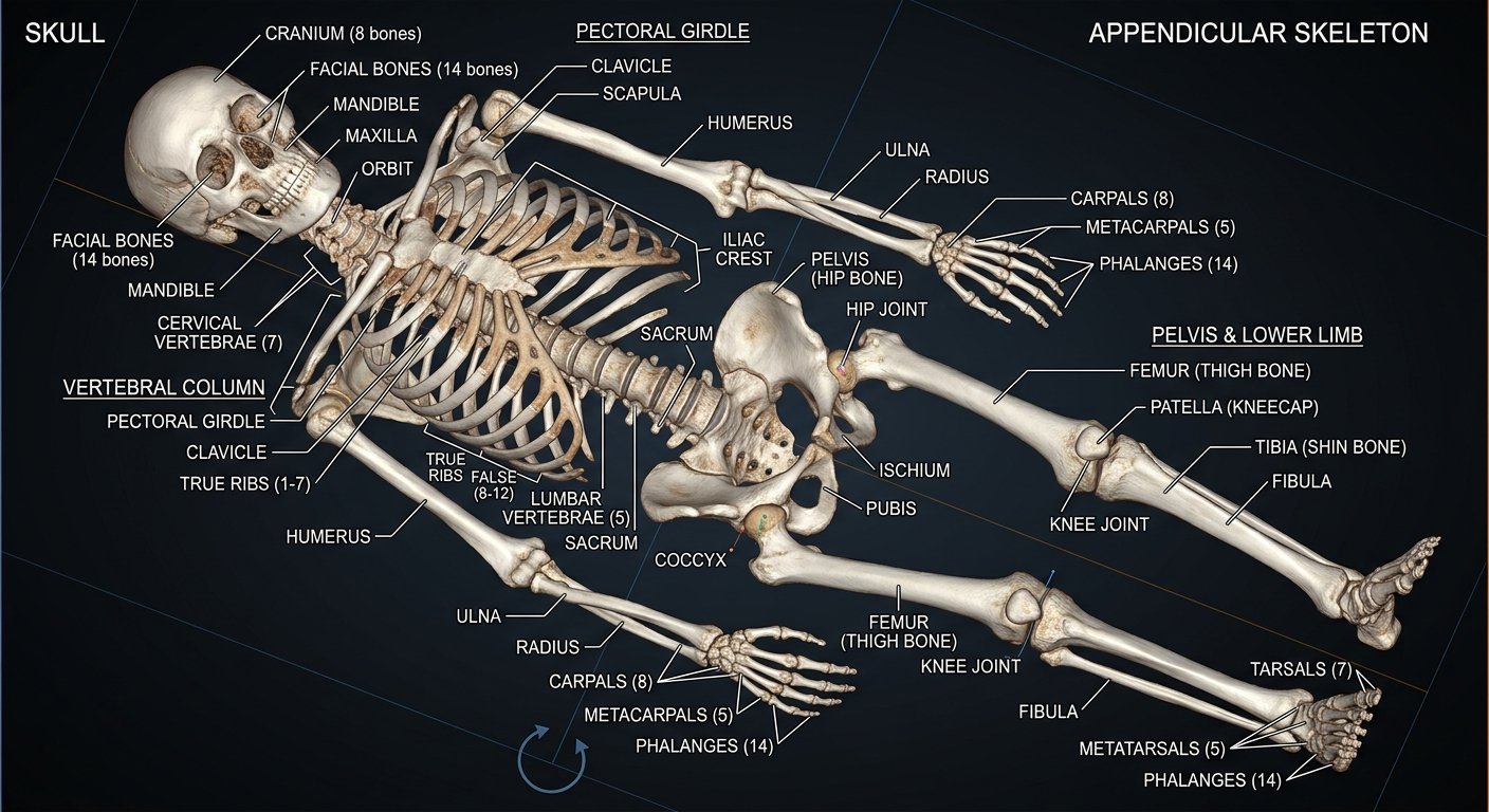
An adult human skeleton is generally described as having 206 bones. The skeleton is divided into two major sections: the axial skeleton and the appendicular skeleton
What Does a Skeleton X-Ray Show?
A full-body skeletal X-ray can demonstrate the major bones and their overall arrangement.
Depending on the image and projection, you may be able to identify:
- Skull
- Jaw
- Clavicles
- Shoulder blades
- Rib cage
- Sternum
- Spine
- Pelvis
- Humerus
- Radius
- Ulna
- Wrist and hand bones
- Femur
- Patella
- Tibia
- Fibula
- Ankle and foot bones
X-rays are particularly useful for showing bone because bone absorbs more X-ray radiation than many of the surrounding soft tissues.
The Two Main Divisions of the Skeleton
1. Axial Skeleton
The axial skeleton forms the central axis of the body.
It includes the:
- Skull
- Vertebral column
- Ribs
- Sternum
- Associated bones such as the hyoid and auditory ossicles
The adult axial skeleton contains about 80 bones.
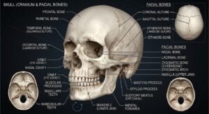
Skull
The skull protects the brain and forms much of the structure of the head and face.
It includes cranial and facial bones such as the frontal, parietal, temporal, occipital, sphenoid, ethmoid, maxilla, zygomatic bones, nasal bones and mandible.
Vertebral Column
The vertebral column, or spine, supports the upper body and surrounds the spinal cord.
It is divided into:
- Cervical vertebrae
- Thoracic vertebrae
- Lumbar vertebrae
- Sacrum
- Coccyx
Rib Cage
The rib cage consists primarily of the ribs and sternum. It forms a protective framework around organs within the chest, including the heart and lungs.
2. Appendicular Skeleton
The appendicular skeleton includes the bones of the limbs and the structures that connect the limbs to the axial skeleton.
It includes:
- Shoulder girdles
- Upper limbs
- Pelvic girdle
- Lower limbs
The appendicular skeleton contains about 126 bones.
Upper Limb Bones
The upper limb is designed for a wide range of movement.
Major bones include:
Clavicle
The clavicle, commonly called the collarbone, connects the shoulder region to the central skeleton.
Scapula
The scapula, or shoulder blade, is a flat bone located at the back of the shoulder.
Humerus
The humerus is the long bone of the upper arm.
Radius and Ulna
The radius and ulna are the two bones of the forearm.
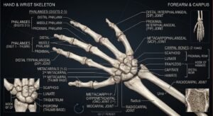
Hand
The hand contains:
- Carpals
- Metacarpals
- Phalanges
Together, these bones allow the hand to perform complex movements.
Lower Limb Bones
The lower limbs are primarily responsible for supporting body weight and producing movement during standing, walking and running.
Femur
The femur is the long bone of the thigh and is the largest bone in the human body.
Patella
The patella, or kneecap, is located at the front of the knee and is a sesamoid bone.
Tibia
The tibia is the larger weight-bearing bone of the lower leg.
Fibula
The fibula is the thinner bone running alongside the tibia.
Foot
The foot contains:
- Tarsals
- Metatarsals
- Phalanges
These bones help provide support, balance and movement.
How Many Bones Are in the Human Skeleton?
The commonly accepted number for an adult human skeleton is 206 bones. However, the exact number can vary because some small accessory bones may be present and because certain bones are formed by fusion during development.
At birth, humans have more separate bones, and some fuse together as the body develops.
What Are the Main Functions of the Skeleton?
The skeletal system performs several important functions.
Support
Bones provide the structural framework that supports the body and helps maintain its shape.
Protection
Bones protect important organs.
For example:
- Skull → protects the brain
- Vertebral column → protects the spinal cord
- Rib cage → helps protect the heart and lungs
- Pelvis → helps protect organs within the pelvic region
Movement
Bones work with muscles and joints to create movement.
Muscles attach to bones through tendons, while joints allow controlled movement between bones.
Mineral Storage
Bones act as an important reservoir for minerals, particularly calcium and phosphorus.
Blood Cell Production
Red bone marrow in certain bones produces blood cells, a process called hematopoiesis.
Types of Bones
Bones can be classified according to their shape.
Long Bones
Long bones are longer than they are wide.
Examples:
- Femur
- Humerus
- Tibia
- Radius
Short Bones
Short bones provide stability and support.
Examples include many of the bones of the wrist and ankle.
Flat Bones
Flat bones often provide protection and broad surfaces for muscle attachment.
Examples include:
- Skull bones
- Ribs
- Sternum
- Scapula
Irregular Bones
These bones have complex shapes.
Examples include:
- Vertebrae
- Some facial bones
Sesamoid Bones
Sesamoid bones develop within certain tendons.
The patella is the largest and most familiar example.
What Can You See in a Skeleton X-Ray?
A skeletal X-ray can make many bones appear clearly because bone absorbs X-rays strongly compared with surrounding soft tissues.
In a full-body skeletal image, you can visually follow the body’s framework from the skull down through the spine, rib cage, pelvis and limbs.
The X-ray can therefore be an excellent visual tool for understanding how individual bones are arranged within the human body.
How Does an X-Ray Create the Image?
During an X-ray examination, X-rays pass through the body toward a detector.
Different tissues absorb different amounts of radiation.
Dense structures such as bone absorb more X-rays and therefore appear relatively bright on the resulting image, while areas where more X-rays pass through appear darker.
The result is a two-dimensional image representing the structures captured by that particular projection.
Skeleton X-Ray vs CT Scan
A conventional X-ray produces a two-dimensional projection.
A CT scan also uses X-rays, but a computer processes multiple measurements to create cross-sectional images and detailed views of internal structures.
Therefore, X-rays and CT scans have different strengths and are selected according to the clinical question.
Interesting Facts About the Human Skeleton
- An adult human skeleton is generally described as containing 206 bones.
- The skeleton is divided into axial and appendicular sections.
- The femur is the longest and one of the strongest bones in the human body.
- The smallest bones are the auditory ossicles inside the middle ear.
- Bones are living tissues that continuously remodel.
- Bone marrow has an important role in blood-cell production.
- Bones store minerals such as calcium and phosphorus.
- The skeleton works together with muscles, tendons, ligaments and joints to produce movement.
Quick Information
| Feature | Details |
|---|---|
| Subject | Human Body Skeleton |
| Category | Human Body |
| Adult bones | Approximately 206 |
| Main divisions | Axial and appendicular |
| Imaging method | X-ray radiography |
| Main functions | Support, protection, movement |
| Other functions | Mineral storage and blood-cell production |
| Related imaging | CT scan |
Explore the Human Skeleton
The human skeleton is much more than a collection of individual bones. It is an integrated structural system that supports the body, protects vital organs, stores minerals and works with muscles to make movement possible.
The X-ray image above provides a visual way to explore this framework from the skull and spine to the hands, pelvis and feet.
Explore each region individually on TECH X-RAY to learn what every major bone does and how it fits into the human body.
Important Note
This article is intended for educational and informational purposes. An X-ray image used for medical diagnosis should be interpreted by a qualified healthcare professional.
Quick Information
Subject: Human Body Skeleton
Category: Human Body
Published: August 27, 2026
Related X-Rays